


If you happen to be on the main Interstate between the University of California, Davis, near Sacramento, and the Golden Gate Fields racetrack, perched on the San Francisco shoreline, and spy an anxious-looking driver at the helm of a horse trailer, chances are you’re in bumping distance to one of two different things of great value.
The first is of the four-legged Thoroughbred variety going from or to the Bay Area facility.
The other is of the one-legged diagnostic variety — more specifically, the $600,000 Longmile Positron Emission Tomography (MILE-PET) Scan machine.
“In the beginning, there was some discussion about how best to move it,” said Mathieu Spriet, associate professor of Surgical and Radiological Sciences at UC Davis, whose brainchild the aforementioned machine is.
“In the end, we went the easy way — in the back of a two-horse trailer,” he added, betraying what might be an approach to home improvements. “We push it up into the trailer, tie it really tight and take it on the I-80. It works really well.”
Reliable technology
As seems to be the fashion nowadays, the absurd rarely appears without the sublime, and in this case this portable diagnostic tool couldn’t be more noble in its intent if it was tethered to the back of a donkey and shunted towards Jerusalem.
That’s because it has the ability to not only catch in racehorses the sorts of brewing injuries that can turn quickly catastrophic but also to detect them earlier than other commonly used pieces of diagnostic wizardry.
In other words, a tool with an answer to one of the biggest questions plaguing the sport.
When Spriet and I spoke, the PET unit had been used at Golden Gate Fields for just over a month, taken there via humble transit once a week. But this same machine has been part of the Santa Anita furniture for over a year and a half, during which time more than 200 horses have been scanned.
“What I’ve learned we can come up with some very specific information for each fetlock, and we can follow that over time,” said Spriet about a procedure that involves injecting a radioactive tracer into the body, which will highlight under imaging abnormal areas in the bone.
More pointedly, “I’m just really happy and excited that we have a reliable technology,” he said, tipping his hat to the weight of expectation hanging over this project.
What it is
Indeed, it was back in 2015 when the first horse was imaged using a PET unit at UC Davis — a global first. That scan, however, was performed using a cumbersome machine developed to image the human brain.
Because the procedure required the horse to be supine and anaesthetized, the potential for PET in the equine world was fairly limited, with all sorts of nasty problems that can arise as horses emerge from their drunken stupor.
What was needed, therefore, was a tool that could be used on a horse stood upright and conscious.
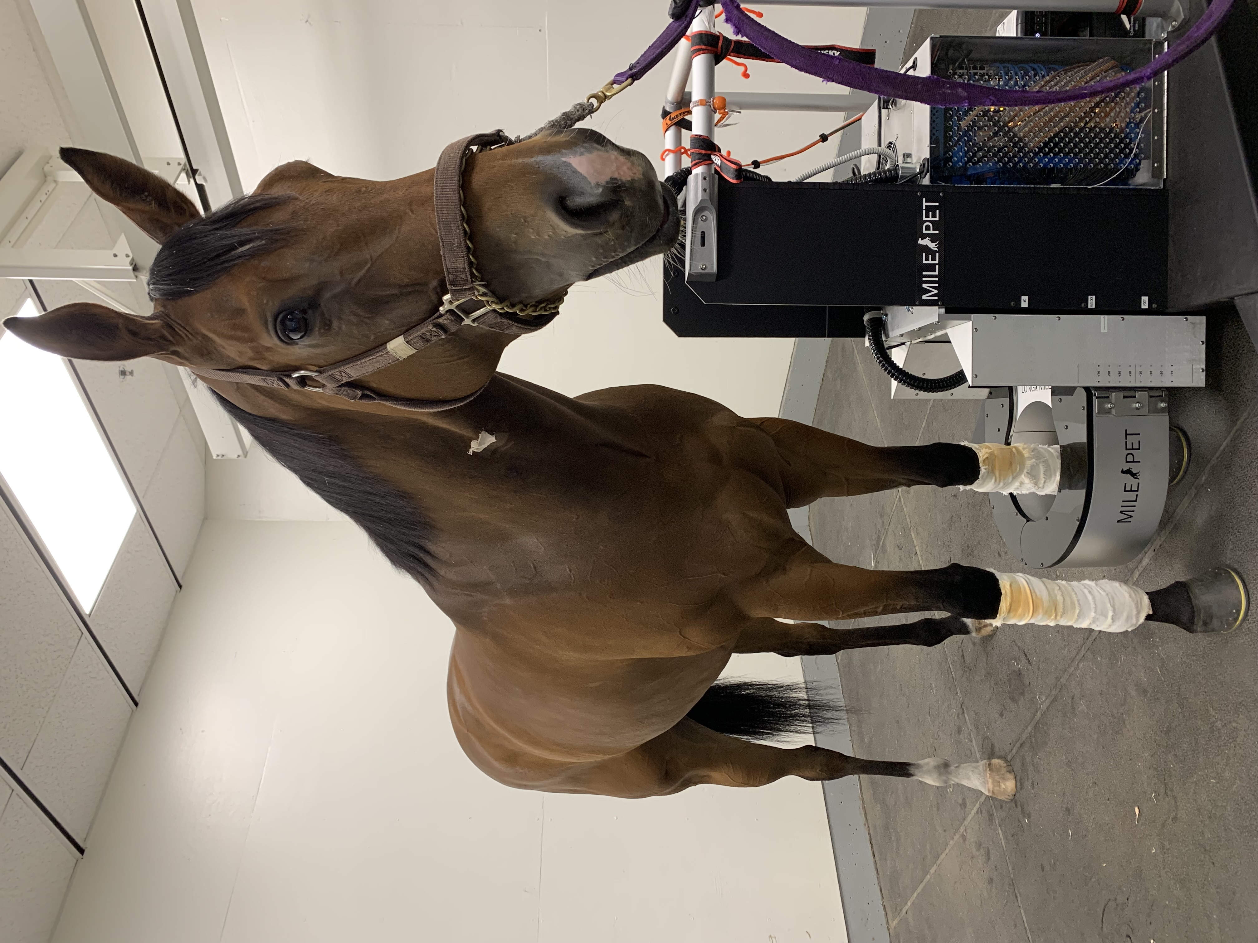
And so Spriet spent the intervening years developing the MILE-PET machine, which resembles a basketball hoop hovering above the ground which can be maneuvered up and down, with a quick-release mechanism to swiftly free any fractious patient.
The machine has been designed to focus on the fetlock, which, given the joint’s size and anatomical complexity, along with the tremendous forces being placed in on it with every race-pace stride, is as close as you’re going to come to an equine version of an Achilles’ heel.
Indeed, experts say that 50-60 percent of catastrophic injuries in racehorses occur at the fetlock joint.
But the potentially revolutionary aspect of the PET is its specificity, especially when it comes to the two delicate little gremlins called the proximal sesamoid bones sitting at the back of the fetlock joint.
Study
Last December, Spriet publicly presented the findings from one year of PET usage at Santa Anita, involving 188 separate scans from 108 different horses.
From this batch of PET scans, the most common problem hotspot was the condyle region of the cannon bone, part of the fetlock joint, and an area notoriously vulnerable to injury.
More importantly, the next most common site of brewing trouble concerned the proximal sesamoid bones. Why?
The PET is the only diagnostic tool currently available with the sensitivity necessary to reliably determine problems in the sesamoids early enough to prevent a potentially catastrophic injury from happening, said Spriet.
“They’re under a lot of stress,” he said, pointing to the competing forces at play on the sesamoids through the loading of the limb and the stresses from the tendons tugging on them.
Indeed, because of their size, these tiny bones are unable to handle the same kinds of forces for as long as other susceptible parts of the horse’s skeleton.
“It takes less pathology to blow up a sesamoid than to blow up a condyle,” said Spriet about the diagnostic window of opportunity. “We really don’t have that much time to catch sesamoid injury. If it progresses too much, it’s likely it will blow.”
As an indication of just how susceptible these bones are to injury, just turn to the benighted winter of discontent at Santa Anita two years ago, when the track came under a spotlight for a rash of fatalities.
A subsequent CHRB report into the 22 equine deaths that resulted from catastrophic musculoskeletal injuries during that period found that 19 of them had sesamoid bone fractures.
“That’s why sesamoid injuries are more concerning,” Spriet added.
Findings
Spriet said that, when it came time to road-test his bespoke PET unit, he was worried the resulting scans would show little variation — in other words, that the machine would show the same things on all racehorses in training.
He needn’t have worried.
“Looking at a lot of different horses, we gain a lot of information very specific to each,” he said, showing me eight slides from eight different horses’ fetlocks, the speckles of cherry red and bright yellow splattered differently from slide to slide.
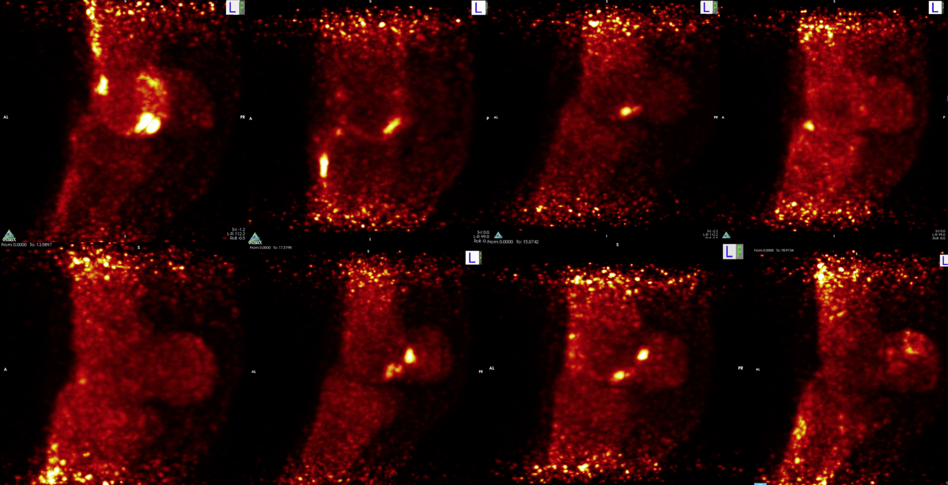
At the same time, certain informative patterns are emerging — in particular, the way in which injuries are repetitive rather than isolated.
“It’s not like, ‘oh, we rested this one because of a condyle [problem]. We brought him back and he blew a sesamoid.’ That’s not how it works,” Spriet explained.
“The one with a hot sesamoid is at risk of heating up the same sesamoid again. The one with a condyle issue, that condyle is at risk of coming back with high intensity training,” he added, describing it as a roadmap of injury for each horse.
“It means that each horse is going to have its own weakness, and we can recognize that and follow that over time.”
As such, Spriet envisages a future whereby researchers, with enough data at their fingertips, might be able to identify trends in injury that correspond with their pedigrees.
MRI
The advent at Santa Anita of an on-site PET machine coincided with that of a standing magnetic resonance imaging (MRI) unit, brought to the track in January last year.
In contrast to PET imaging — which observes activity at the molecular level — the MRI unit harnesses the body’s natural magnetic properties to provide “morphological” information, which are those related to the size, shape or density of the Musculo skeleton.
While the PET has proven hyper-sensitive to changes in the sesamoids, the MRI has been adept at recognizing brewing trouble in the condylar region of the fetlock, said Southern California-based private veterinarian Ryan Carpenter.
“I think MRI will pick up changes in the condyle before the PET,” said Carpenter. “But I think the great thing about PET is it’s a three-dimensional image. It’s very sensitive and specific for regions.”
More broadly, Carpenter said that as PET and MRI become a more standard part of the veterinarian nomenclature, and as the science behind the standing computed tomography (CT) unit — what he calls the “next modality to come around” — continues to evolve, diagnosticianns are only going get better at targeting different tools for specific injuries.
We didn’t get here by accident.
“A couple years ago, we realized imaging was going to be a really important thing for the future,” Carpenter said.
“A lot of research institutes poured a lot of money into imaging,” he added. “It takes a long time to do good research and get good results, and I think what we’re going to see this year and next year is the fruit of all that labor.”
Future
Spriet and his cohorts in the brave new world of the standing PET — developed with funding from a variety of sources, including the Grayson-Jockey Club Research Foundation, The Stronach Group, the Dolly Green Research Foundation, and UC Davis Center for Equine Health — have already embarked upon a fishing expedition looking at horses showing no visible sign of lameness.
To do this, researchers randomly selected horses that raced at Santa Anita or Golden Gate Fields and finished among the leaders, the idea being those horses that performed creditably are the ones least likely to be suffering ailments.
Of the 40 or so horses studied thus far, said Spriet, about half showed no visible brewing problems. “In almost the other half, we saw changes that I’d consider mild, meaning there are no real issues with these horses,” he added.
Nevertheless, about four of the fetlocks studied in this population had problems severe enough to likely require veterinary intervention, even time off, he said.
Later this summer, Spriet will be lending a helping hand to researchers at the University of Pennsylvania as they scan horses housed at Parx Racing.
As Spriet sees it, this is an opportunity to see how different facilities and training regimens cause different sorts of cyclical wear-and-tear on the fetlock joint. “That’s going to be really interesting to compare California racetracks with the East Coast,” he said.
Asked to make a prediction about what they might find, Spriet said the East Coast tends to see slightly fewer sesamoid injuries and more condylar fractures as compared to California. Dogmatism, however, isn’t part of his play-book.
“I’ve learned a lot,” he said. “But I’ve a lot more to learn.”